Steinmetz data set, Moniz 2017-05-15.
So I'm analyzing data from this paper https://www.nature.com/articles/s41586-019-1787-x The dataset is available here https://figshare.com/articles/steinmetz/9598406. I worked on it during Neuromatch Academy and now I've been playing around with it, experimenting, injecting spikes into simulations and stuff. You can check out some of my experiments in these repository https://github.com/mariakesa/NeuroPixelsDataAnalysis
So there are about 30 mice and all the Neuropixel probes are in different locations in the brain. So I made a script to print out which brain areas are recorded from in each mouse. I've also started getting interested in connectivity between areas and in brain anatomy so I'm using the Allen Brain Explorer https://connectivity.brain-map.org/static/brainexplorer to visualize the areas. It's got a cool 3D interface. Nothing like seeing it in 3D:-)
So I thought I'd start documenting the mice in this blog for my own reference and posting pictures from the atlas. I thought I'd actually get to know the mice:-) Plus, I want to write about the brain areas. I basically search for the brain area in Neuron:-)
For your reference, here's a nomenclature of brain region abbreviations that I used http://www.mbl.org/abbreviations/abbrev_frame.html
So let's start with not my most favorite scientist (the mice are named after Nobel Prize winners:-D), Moniz. He invented the lobotomy:-D I
I am looking at the 2017-05-15 recording, it looks like a single NP probe was inserted at a particular time.
This mouse has the following brain regions at this time point:
1. APN --Anterior pretectal nucleus (not in the brain region abbreviations reference). There's 185 neurons!
So it's a midbrain structure, which is forward-most part of the brainstem.
Here's a picture in the A Brain Explorer. Cortex is in light green, APN is in bright green.
Neuron says that APN projects to the lateral posterior nucleus of the thalamus (LP) (in bright green in the picture, APN now in light green), which projects to V1 https://www.cell.com/neuron/fulltext/S0896-6273(21)00283-X 
It's got projections from S1 (primary somatosensory cortex) https://www.cell.com/neuron/fulltext/S0896-6273(11)00682-9
Here's a Wikipedia article that talks about the pretectal area in general https://en.wikipedia.org/wiki/Pretectal_area It says that pretectal neurons receive inputs from the retina and are involved in mediating responses to acute changes in ambient light, such as the pupillary light reflex and the optokinetic reflex. All pretectal nuclei, except for the ON, project to nuclei in the thalamus, subthalamus, superior colliculus, reticular formation, pons, and inferior olive. It's part of the subcortical visual system.
2. CA1-- Ammon's horn, field CA1.
There's a lot of material on CA1, it's a very famous area. From Wikipedia https://en.wikipedia.org/wiki/Hippocampus_proper CA1 is the first region in the hippocampal circuit, from which a major output pathway goes to layer V of the entorhinal cortex. Another significant output is to the subiculum.
This thing represents the environment via place cells! https://www.cell.com/neuron/fulltext/S0896-6273(20)30858-8 -- it's a really cool paper by the way:-) And these cells ensembles have memory replays! https://www.cell.com/neuron/fulltext/S0896-6273(17)30857-7
3. DG-- Dentate Gyrus.
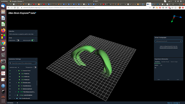
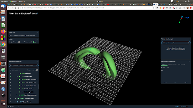
Here I've plotted it along with CA1, the CA1 is the external green layer, DG is the internal green layer (I don't know how to change the colors!).
From Wikipedia: The dentate gyrus (DG) is part of the hippocampal formation in the temporal lobe of the brain that includes the hippocampus and the subiculum. The dentate gyrus is part of the hippocampal trisynaptic circuit and is thought to contribute to the formation of new episodic memories,[1][2] the spontaneous exploration of novel environments[2] and other functions.
This is a site of pattern separation, memory discrimination. This paper has the most hypnotic calcium imaging videos https://www.cell.com/neuron/fulltext/S0896-6273(20)30271-3 :-D
4. LP-- Lateral posterior nuclues of the thalamus.
Here in pink along with CA1 and DG.
5. POL-- Posterior limiting nucleus of the thalamus.
In pink! along with LP and the hippocampal areas:-)
Couldn't find anything on it.
6. SUB-- Subiculum
In dark green!
It is hypothesized to be involved in working memory and CA1 projects to it.
7.VISam-- Anteromedial visual area.
In blue. Visual area, V2. Couldn't find anything specific on it.
In general, Wikipedia on V2: Cells are tuned to simple properties such as orientation, spatial frequency, and color. The responses of many V2 neurons are also modulated by more complex properties, such as the orientation of illusory contours,[24][25] binocular disparity,[26] and whether the stimulus is part of the figure or the ground.[27][28] Recent research has shown that V2 cells show a small amount of attentional modulation (more than V1, less than V4), are tuned for moderately complex patterns, and may be driven by multiple orientations at different subregions within a single receptive field.
8.VISpm-- Posteromedial visual area.
In blue, posterior to VISam.
Part of V2. I couldn't find anything specific on it!
As a summary, this data set contained neurons from the midbrain (brainstem) involved in light reflexes, hippocampus (place fields, pattern separation regions), visual thalamus (with retinotopy) and V2, in which cells are tuned to orientation and color and also more complex patterns and show a bit of attentional modulation.


Kommentaarid
Postita kommentaar